Tests and treatments
- Accepting New Patients
- Most Insurance Plans Accepted
- Member of the Chamber of Commerce
(713) 258-6111
Tests and Treatments

Examination
The examination consists in the doctor examining the chest and listening to the chest with a little microphone called stethoscope to try and spot some clues which might be able to establish the cause of the symptom or complaint. This is the way most heart murmurs are found.
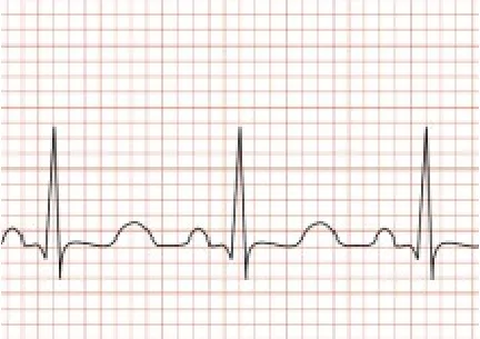
ECG
The ECG consists in the recording of the electrical activity of the heart on paper. ECG is performed by applying 10 small stickers to the child skin, 6 on the chest and one each in each arm and leg. The stickers have the purpose of picking up the heart’s electrical signals. The ECG takes less than 5 minutes to complete and is completely pain free. After the test the stickers are easily removed from the skin and the test result is available immediately.
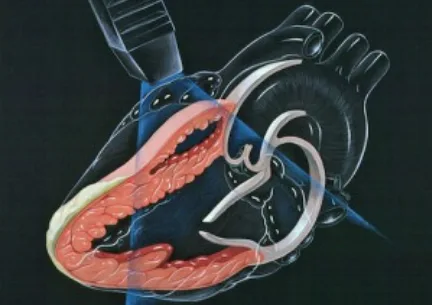
Echocardiogram
The echocardiogram is a non-invasive test based on ultrasounds which allows visualizing the cardiac structures and the blood flow in the heart and the large chest veins and arteries. The ultrasounds are produced by a little microphone and some jelly is applied on the chest and on the stomach to improve the quality of the images. With an echocardiogram it is possible to make a complete diagnosis in almost all cases of congenital heart defects (CHD). As the technique is completely non invasive and pain free this is ideally used in children. The test takes around 15 to 20 minutes and the result is generally available immadiately.
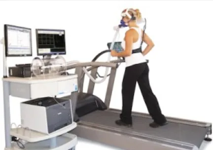
Exercise test
The exercise test consists of the controlled measurement of a child heart, lungs, circulation and muscles to exercise. The test is used to:
1) investigate the causes of chest pain, and palpitations
2) investigate the causes of fainting during exercise
3) investigate the causes of breathlesness during exercise, including asthma
4) investigate the causes of easy fatigue
5) assess fitness level in children before the start of intense exercise
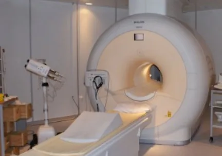
Cardiac CT/MRI
Cardiac magnetic resonance imaging (MRI) is a safe and completely non invasive test that creates detailed pictures of your organs and tissues. MRI uses radio waves, magnets, a special dye, and a computer to create pictures of your organs and blood vessels. Unlike chest X-ray and computed tomography (CT), MRI does not use any ionizing ratiation and carries no risk of causing cancer. Cardiac MRI produces both still and moving images of the heart and major blood vessels. Doctors use cardiac MRI to get pictures of the heart while beating and to look at how it is made and functions. These pictures can help them decide the best way to treat children with heart problems. The test takes around 45 minutes to complete and a small venous line has to placed in order to allow for dye injection. Younger children require a general anaesthetic in order to remain completely still for the duration of the test.
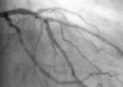
Cardiac catheterization
Cardiac catheterization is a medical procedure used to diagnose and treat some congenital heart defects and conditions in babies and children. A long, thin, flexible tube called a catheter is put trough a blood vessel in the groin (upper thigh) or neck and threaded to the heart. Trough the catheter, doctors can do some diagnostic tests (like measure pressure in the cardiac chambers) and carry out treatments on the heart. For example the doctor may put a special type of dye (contrast) in the catheter. As a result the dye will flow trough the bloodstream to the heart. Then, the doctor will take X-ray pictures of the heart known as angiogram. If any problem is seen it can be sometimes treated at the same time. This test will require a general anaesthetic to be performed. The test generally takes around 2 hours to be performed and most children can be discharged home on the same day of treatment.
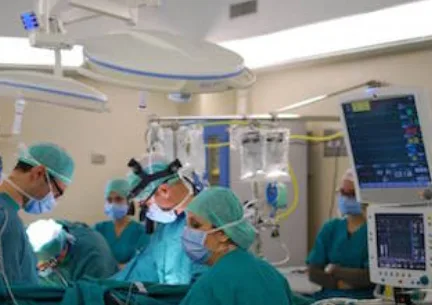
Cardiac Surgery
Heart surgery in children is undertaken to repair heart defects a child is born with (congenital heart defects or CHD) and heart diseases a child gets after birth that needs surgery. The surgery is needed for the child weel being.
There are many kinds of heart defects. Some cardiac defects are minor whereas others are more serious. Defects can occur inside the heart or in the large blood vessels outside the heart. Some heart defects may require for surgery to take place right after the baby is born. For others, your child may be able to safely wait for months or years to have surgery. In some types of cardiac defects the surgeons have to stop the heart and a heart-lung machine has to be used in order to support the blood flow and baby functions while the surgeons are operating.
When the heart and lung machine has to used, an incision is made through the breastbone (sternum) while the child is under general anaesthesia (the child is unconscious and does not feel pain). For the repair of some heart defects, the incision has to be made on the side (left or right) of the chest, between the ribs. Once the surgery is completed the child will be transferred to the cardiac intensive care unit and as she makes progress she is prepared for discharge. The length of time to be spent in hospital after a cardiac operation can be quite variable but most children can be discharged within 7-10 days from the operation. After the operation, a specifically tailored follow-up schedule will be organized to suit the child and the family needs.
New Patient Paperwork
Please print out your paperwork and bring it to your appointment at SW Houston Cardiology. If your insurance requires authorization, please be sure to have that information sent to our office or bring it with you to your appointment, along with any medical records you may have. Please also make sure to bring your insurance cards, photo ID, and medication list.
Thank you, and we look forward to meeting you! Contact us today with any questions.
Call to Schedule Your Appointment!
Most insurance plans accepted
(713) 258-6111
